Difference between revisions of "Microbe Mission"
m |
|||
| (18 intermediate revisions by 5 users not shown) | |||
| Line 1: | Line 1: | ||
{{EventLinksBox | {{EventLinksBox | ||
| − | |active= | + | |active= |
|type=Life Science | |type=Life Science | ||
|cat=Lab | |cat=Lab | ||
| Line 16: | Line 16: | ||
|C Champion=[[Mason High School]] | |C Champion=[[Mason High School]] | ||
}} | }} | ||
| − | In '''Microbe Mission''', teams will answer questions, solve problems, and analyze data pertaining to microbes and [[Microscope|microscopes]]. The event was run as a trial event before being made an official event in [[2011]] and [[2012]], and has returned for the [[2017]] and [[2018]] season. | + | In '''Microbe Mission''', teams will answer questions, solve problems, and analyze data pertaining to microbes and [[Microscope|microscopes]]. The event was run as a trial event before being made an official event in [[2011]] and [[2012]], and has returned for the [[2017]] and [[2018]] season. It is recommended that competitors also view the [[Microbe Mission Diseases List|Microbe Mission Diseases List wiki page]]. |
If you are interested in helping by improving the Microbe Mission page(s), see [[Microbe Mission/Improvement|this page]]. | If you are interested in helping by improving the Microbe Mission page(s), see [[Microbe Mission/Improvement|this page]]. | ||
==Event Overview== | ==Event Overview== | ||
| − | + | Each team will be tested on topics relating to microbiology, ranging from different types of [[Microscope|microscopes]] and microbes to learning about cell structure and uses for microbes. Teams must also be able to read a dichotomous key and understand the principles of microscopy. | |
===The Stations=== | ===The Stations=== | ||
| − | If there are stations, there will be 8-20 of them. They will be marked with | + | If there are stations, there will be 8-20 of them. They will be marked with Roman numerals (I, II, III...) or they will be numbered (1, 2, 3...). There will be sections in the test corresponding to each of the stations with questions (the format of which is decided by the tester and can vary widely from tester to tester). Students typically have a time limit at stations (i.e. 5 minutes per station, then rotate). |
===The Test=== | ===The Test=== | ||
| − | The test will pages/sections corresponding to the individual stations (if there aren't stations then it will be a normal test). It will have blank lines for | + | The test will pages/sections corresponding to the individual stations (if there aren't stations then it will be a normal test). It will have blank lines for teams to record their answer. There will be no questions/diagrams in the packet, so all work must be done at the corresponding station. At some competitions, the questions will be at the station. All answers must be recorded in the packet. Spelling does count in the packet. Points may also be taken away if the packet is not neat or legible. As teams record their answers, they should ensure that they are recording on the right page/section/question. This may save time and effort. |
| − | Please note that there may be lines for | + | Please note that there may be lines for team name, team number, or the participants' names '''on each page'''. Teams should ensure their information is present on each page. Points can be deducted from teams who fail to do so. |
===Materials=== | ===Materials=== | ||
| Line 37: | Line 37: | ||
==Preparing for This Event== | ==Preparing for This Event== | ||
| − | + | Making a binder can be an essential step in preparing for this event. Even though binders are not permitting during testing, it's a great way to keep all information in the same place for studying or review. | |
| − | With | + | With a notes page, include pictures and charts concerning the tested topics. Also, it is crucial to remember how the page is laid out. In a timed test, locating information on the sheet quickly is a pivotal skill. |
| − | + | An AP Biology textbook will help tremendously with learning terms and concepts. | |
| − | + | Practice allows one to familiarize with a variety of topics for the type of questions and how they are presented can vary widely from test to test. Practice tests are extremely helpful in preparing for this event. | |
'''Tips for Making the Note Sheet''' | '''Tips for Making the Note Sheet''' | ||
| − | *Use as small of a font as | + | *Use as small of a font as possible. However, it is advised to '''keep it readable'''. There's no point in having volumes of information if it is impossible to interpret. |
*Make your own diagrams, either by hand or with an image manipulation program (paint). | *Make your own diagrams, either by hand or with an image manipulation program (paint). | ||
| − | *Color code. Use a different (readable) color for notes on each system. This will make things easy to find | + | *Color code. Use a different (readable) color for notes on each system. This will make things easy to find on competition day. Also, color-coded diagrams tend to increase one's efficiency in interpretation. (as seen in the picture above). It's much easier to find a bright orange muscle than one outlined lightly in black. Keep the coding consistent so that by the end of the season you automatically associate a color with a type of information (ex: pink = muscles; blue = respiratory; green = endocrine and etc.) Highlighting may save a lot of time at the competition. |
| − | * | + | *Additional notes can be handwritten in places where the printer might not be able to print. This is time-consuming but well worth the time spent. |
| − | *Source-check before doing anything. | + | *Source-check before doing anything. This will allow one to avoid putting incorrect information on a note sheet, saving both time and energy in having to correct mistakes in the future. |
| − | *Use space efficiently by prioritizing. Include the | + | *Use space efficiently by prioritizing. Include the material that is hardest to understand or remember. Extra information can be added later if there is still room available. |
| − | *Use charts, as they are life-savers. | + | *Use charts, as they are life-savers. Charts can be personally made to include specific information - the simple act of making a chart can help tremendously to maximize space. By personally developing a chart, one can also gain a better understanding of the material. |
| − | *Laser printers are recommended | + | *Laser printers are recommended for smaller font sizes. Font sizes can be reduced manually if you treat text like a picture (by typing it onto an image manipulation program and then shrinking the image), though this may reduce the readability of the notes. |
| − | * | + | *Communication among partners is vital in every event. It is important to ensure that both partners understand and remember the layout of the sheet. This will allow both members to test as efficiently as possible. |
| − | *A | + | *A recommended font for Microbe Mission notes (or any other note-based event) is BenchNine, in either 6 or 7 font size, depending on which both partners can read more easily. |
==Types of Microbes== | ==Types of Microbes== | ||
| Line 68: | Line 68: | ||
Some viruses, known as bacteriophages, infect bacteria. Their appearance is often compared to that of an alien landing pod. Typically, their genome is composed of DNA rather than the RNA of retroviruses. Other viruses, most famously Sputnik, infect other viruses. These are known as virophages. | Some viruses, known as bacteriophages, infect bacteria. Their appearance is often compared to that of an alien landing pod. Typically, their genome is composed of DNA rather than the RNA of retroviruses. Other viruses, most famously Sputnik, infect other viruses. These are known as virophages. | ||
| − | Viruses can be caused either lytic or lysogenic infections. In a lytic infection, the virus injects its genome into the host cell, which cannot differentiate between viral DNA and its own DNA. The cell begins to make mRNA from the viral DNA, which is then made into viral proteins that destroy the cell's DNA. When the cell eventually shuts down, the virus continues to use the cell to replicate. Enough viruses are made to cause the cell to burst, or lyse. Hundreds or thousands of released viruses then go on to infect other cells. | + | Viruses can be caused by either lytic or lysogenic infections. In a lytic infection, the virus injects its genome into the host cell, which cannot differentiate between viral DNA and its own DNA. The cell begins to make mRNA from the viral DNA, which is then made into viral proteins that destroy the cell's DNA. When the cell eventually shuts down, the virus continues to use the cell to replicate. Enough viruses are made to cause the cell to burst, or lyse. Hundreds or thousands of released viruses then go on to infect other cells. |
In a lysogenic infection, a virus integrates its DNA into the host cell's DNA. This viral DNA is known as a prophage. The prophage remains dormant in the cell's DNA for several generations before becoming active, leaving the cell's DNA, and directing the synthesis of new viral proteins. HIV, which causes AIDS, is a lysogenic virus. | In a lysogenic infection, a virus integrates its DNA into the host cell's DNA. This viral DNA is known as a prophage. The prophage remains dormant in the cell's DNA for several generations before becoming active, leaving the cell's DNA, and directing the synthesis of new viral proteins. HIV, which causes AIDS, is a lysogenic virus. | ||
| Line 74: | Line 74: | ||
===Cellular Microbes=== | ===Cellular Microbes=== | ||
| − | Cellular microbes are microbes that are made up of cells. There are two main types of cellular microbes; [[Prokaryote|prokaryotes]] and [[Eukaryote|eukaryotes]]. Prokaryotes differ from eukaryotes with their lack of nuclei and membrane bound organelles. | + | Cellular microbes are microbes that are made up of cells. There are two main types of cellular microbes; [[Prokaryote|prokaryotes]] and [[Eukaryote|eukaryotes]]. Prokaryotes differ from eukaryotes with their lack of nuclei and membrane-bound organelles. |
====Bacteria==== | ====Bacteria==== | ||
| Line 92: | Line 92: | ||
====Protists==== | ====Protists==== | ||
| − | Protists are [[eukaryote|eukaryotic]] but do not have specialized tissues. Algal protists are similar to plants and can go through photosynthesis, but do not have cuticles that prevent water loss. As a result algal protists must live in water. Animal like protists are called protozoa and are eukaryotic and heterotrophic. These protists consume other protists and bacteria for food. Some have two nuclei: the macronucleus and the micronucleus. Many move with cilia, flagella, or pseudopodia (in the case of amoebae). They also have complex life cycles. For example, they may exist in a '''trophozoite''', or feeding, form. They can also change into a dormant form known as a '''cyst''', which can help in reproduction. | + | Protists are [[eukaryote|eukaryotic]] but do not have specialized tissues. Algal protists are similar to plants and can go through photosynthesis, but do not have cuticles that prevent water loss. As a result, algal protists must live in water. Animal-like protists are called protozoa and are eukaryotic and heterotrophic. These protists consume other protists and bacteria for food. Some have two nuclei: the macronucleus and the micronucleus. Many move with cilia, flagella, or pseudopodia (in the case of amoebae). They also have complex life cycles. For example, they may exist in a '''trophozoite''', or feeding, form. They can also change into a dormant form known as a '''cyst''', which can help in reproduction. |
==Endosymbiotic Theory== | ==Endosymbiotic Theory== | ||
| − | Championed by Lynn Margulis in the 1960s, the endosymbiotic theory holds that mitochondria and chloroplasts in eukaryotic cells originated from proteobacteria and cyanobacteria, respectively. Evidence for this theory includes that mitochondria and chloroplasts divide through binary fission, not mitosis like the rest of the cell. These organelles, which are the same size as bacteria, also have their own different, circular DNA, their own ribosomes and two membranes. The two membranes have different chemical compositions, with the outer being similar to the eukaryotic plasma membrane and the inner being similar to bacterial membranes. Chloroplasts in some algae have cell walls of peptidoglycan. | + | Championed by Lynn Margulis in the 1960s, the endosymbiotic theory holds that mitochondria and chloroplasts in eukaryotic cells originated from proteobacteria and cyanobacteria, respectively. Evidence for this theory includes that mitochondria and chloroplasts divide through binary fission, not mitosis like the rest of the cell. These organelles, which are the same size as bacteria, also have their own different, circular DNA, their own ribosomes, and two membranes. The two membranes have different chemical compositions, with the outer being similar to the eukaryotic plasma membrane and the inner being similar to bacterial membranes. Chloroplasts in some algae have cell walls of peptidoglycan. |
==Microbial Growth Curve== | ==Microbial Growth Curve== | ||
| Line 113: | Line 113: | ||
'''Decline or Death Phase''' | '''Decline or Death Phase''' | ||
| − | *Unless the microbes have an infinite source of nutrients, the microbes begin to use up all of the surrounding resources and the waste products begin to pile up. This leads to an unfavorable environment which causes the amount of dying cells outnumber the amount of new ones. | + | *Unless the microbes have an infinite source of nutrients, the microbes begin to use up all of the surrounding resources and the waste products begin to pile up. This leads to an unfavorable environment which causes the amount of dying cells to outnumber the amount of new ones. |
==Gram Staining== | ==Gram Staining== | ||
| Line 128: | Line 128: | ||
===Characteristics of Gram-Positive Bacteria=== | ===Characteristics of Gram-Positive Bacteria=== | ||
| − | Typically, Gram-positive bacteria produce exotoxins and are susceptible to phenol disinfectants. They retain the blue-purple color of crystal violet in Gram staining because of their thicker walls of peptidoglycan. Unlike Gram-negative bacteria, they lack the periplasmic space between the cytoplasmic and outer membranes because Gram-positive bacteria lack an outer membrane. Certain types of Gram-positive bacilli, most importantly ''Lactobacilli'' (used in milk and dairy products), cannot form | + | Typically, Gram-positive bacteria produce exotoxins and are susceptible to phenol disinfectants. They retain the blue-purple color of crystal violet in Gram staining because of their thicker walls of peptidoglycan. Unlike Gram-negative bacteria, they lack the periplasmic space between the cytoplasmic and outer membranes because Gram-positive bacteria lack an outer membrane. Certain types of Gram-positive bacilli, most importantly ''Lactobacilli'' (used in milk and dairy products), cannot form spores. |
===Characteristics of Gram-Negative Bacteria=== | ===Characteristics of Gram-Negative Bacteria=== | ||
| Line 134: | Line 134: | ||
==Types of Microscopes== | ==Types of Microscopes== | ||
| + | {{Cleanup|section}} | ||
| + | ''Main article: [[Microscope]]'' | ||
| − | *Optical Microscopes: Optical microscope uses visible light (or UV light in the case of fluorescence microscopy) to sharply magnify the samples. The light rays refract with optical lenses. Dating back to the first microscopes that were invented, it is found that they belonged to this category. Optical microscopes can be further subdivided into several categories: | + | *'''Optical Microscopes''': Optical microscope uses visible light (or UV light in the case of fluorescence microscopy) to sharply magnify the samples. The light rays refract with optical lenses. Dating back to the first microscopes that were invented, it is found that they belonged to this category. Optical microscopes can be further subdivided into several categories: |
| − | *Compound Microscope: The compound microscope is built of two systems of lenses for greater magnification (an objective and an ocular: eyepiece). The utmost useful magnification of a compound microscope is about 1000x. | + | *'''Compound Microscope''': The compound microscope is built of two systems of lenses for greater magnification (an objective and an ocular: eyepiece). The utmost useful magnification of a compound microscope is about 1000x. |
| − | *Stereo Microscope (dissecting microscope): The stereo microscope is an optical microscope which magnifies up to about maximum 100x and provides a 3-dimensional view of the specimen. Stereo microscopes are highly useful for observing opaque objects. | + | *'''Stereo Microscope''' (dissecting microscope): The stereo microscope is an optical microscope which magnifies up to about maximum 100x and provides a 3-dimensional view of the specimen. Stereo microscopes are highly useful for observing opaque objects. |
| − | *Confocal Laser scanning microscope: Unlike compound and stereo microscopes, Confocal Laser scanning microscopes are reserved for research organizations. Such microscopes are able to scan a sample also in depth. A computer can then assemble the data to create a 3D image. | + | *'''Confocal Laser scanning microscope''': Unlike compound and stereo microscopes, Confocal Laser scanning microscopes are reserved for research organizations. Such microscopes are able to scan a sample also in depth. A computer can then assemble the data to create a 3D image. |
| − | *Electron Microscopes:Electron microscopes are the most advanced microscopes used in modern science. The electron microscopes essentially function on the principle of a beam of electrons that strikes any objects that comes to its path to magnify it. Electron microscopes are designed specifically for studying cells and small particles of matter, as wells as large objects. | + | *'''Electron Microscopes''': Electron microscopes are the most advanced microscopes used in modern science. The electron microscopes essentially function on the principle of a beam of electrons that strikes any objects that comes to its path to magnify it. Electron microscopes are designed specifically for studying cells and small particles of matter, as wells as large objects. |
| − | *Scanning Electron Microscope: Scanning Electron Microscope is characterized as a microscope that has lower magnifying power but can provide 3 dimensional viewing of objects. The Scanning Electron Microscope captures the image of the object in black and white after being stained with gold and palladium. | + | *'''Scanning Electron Microscope''': Scanning Electron Microscope is characterized as a microscope that has lower magnifying power but can provide 3-dimensional viewing of objects. The Scanning Electron Microscope captures the image of the object in black and white after being stained with gold and palladium. |
| − | *Reflection Electron Microscope: Reflection electron microscopes are also designed on the principle of electron beams but they are characteristically different from transmission and scanning electron microscopes being that it is built to detect electrons that have been scattered elastically. | + | *'''Reflection Electron Microscope''': Reflection electron microscopes are also designed on the principle of electron beams but they are characteristically different from transmission and scanning electron microscopes being that it is built to detect electrons that have been scattered elastically. |
| − | *X-ray Microscope: An X-ray microscope uses a beam of x-rays to create an unparalleled high resolution 3D image. Due to the small wavelength, the image resolution is higher as compared to optical microscopes. The greatest useful magnification is therefore also higher and it lies between the optical microscopes and electron microscopes. X-ray microscopes hold significant importance in science and research and have got one special advantage over electron microscopes- it allows observing the structure of the living cells. It is adept at slicing together thousands of images to generate a single 3D X-ray image. | + | *'''X-ray Microscope''': An X-ray microscope uses a beam of x-rays to create an unparalleled high-resolution 3D image. Due to the small wavelength, the image resolution is higher as compared to optical microscopes. The greatest useful magnification is therefore also higher and it lies between the optical microscopes and electron microscopes. X-ray microscopes hold significant importance in science and research and have got one special advantage over electron microscopes- it allows observing the structure of the living cells. It is adept at slicing together thousands of images to generate a single 3D X-ray image. |
| − | *Scanning Helium Ion Microscope (SHIM or HeIM): Scanning Helium Ion Microscope is a new imaging technology which uses a beam of Helium ions beams to generate an image. This technology has several advantages over the traditional electron microscopes; one advantage lies in the fact that the sample is left mostly intact (due to the low energy requirements) and that it provides a high resolution. The first commercial systems were released in 2007. | + | *'''Scanning Helium Ion Microscope''' (SHIM or HeIM): Scanning Helium Ion Microscope is a new imaging technology which uses a beam of Helium ions beams to generate an image. This technology has several advantages over the traditional electron microscopes; one advantage lies in the fact that the sample is left mostly intact (due to the low energy requirements) and that it provides a high resolution. The first commercial systems were released in 2007. |
| − | *Scanning acoustic microscope (SAM): Scanning acoustic microscope uses focused sound waves to generate an image. An acoustic microscope has a wide range of applications in materials science to detect small cracks or tensions in materials. The scanning acoustic microscope is a powerful tool which can also be used in biology to study the physical properties of the biological structure and help uncover tensions, stress and elasticity inside the biological structure. | + | *'''Scanning acoustic microscope''' (SAM): Scanning acoustic microscope uses focused sound waves to generate an image. An acoustic microscope has a wide range of applications in materials science to detect small cracks or tensions in materials. The scanning acoustic microscope is a powerful tool which can also be used in biology to study the physical properties of the biological structure and help uncover tensions, stress and elasticity inside the biological structure. |
| − | *Electron Microscopes: Modern electron microscope uses accelerated electrons and can magnify up to 2 million times due to the very small wavelength of high energy electrons. The high energy electrons as a source of illumination are quite tough on the sample being observed. Because of the much shorter wavelength, the electron microscope has a higher resolving power than a light microscope. To reveal the structure of objects, it may initially acquire a long time to completely dehydrate and prepare the specimen. A sleek layer of a metal could be on the other hand used to coat some of the biological specimens for easy observation. | + | *'''Electron Microscopes''': Modern electron microscope uses accelerated electrons and can magnify up to 2 million times due to the very small wavelength of high energy electrons. The high energy electrons as a source of illumination are quite tough on the sample being observed. Because of the much shorter wavelength, the electron microscope has a higher resolving power than a light microscope. To reveal the structure of objects, it may initially acquire a long time to completely dehydrate and prepare the specimen. A sleek layer of a metal could be on the other hand used to coat some of the biological specimens for easy observation. |
| − | *Neutron Microscope: Still under an experimental stage, Neutron microscope generates a high resolution image and may offer better contrast than other forms of microscopy. The new technology would use neutrons instead of beams of light or electrons to generate high resolution images. | + | *'''Neutron Microscope''': Still under an experimental stage, Neutron microscope generates a high-resolution image and may offer better contrast than other forms of microscopy. The new technology would use neutrons instead of beams of light or electrons to generate high-resolution images. |
| − | *Scanning Probe Microscopes: Scanning Probe Microscope helps visualize individual atoms. The image of the atom is computer-generated, however. It provides the researchers an imaging tool for the future where a small tip measures the surface structure of the sample. These specialized microscopes provide high image magnification to observe three dimensional specimens. If an atom projects out of the surface, then a higher electrical current flows through the tip. The amount of current that flows is proportional to the height of the structure. A computer then assembles the position data of the tip. An enhanced 3D image is generated. | + | *'''Scanning Probe Microscopes''': Scanning Probe Microscope helps visualize individual atoms. The image of the atom is computer-generated, however. It provides the researchers with an imaging tool for the future where a small tip measures the surface structure of the sample. These specialized microscopes provide high image magnification to observe three-dimensional specimens. If an atom projects out of the surface, then a higher electrical current flows through the tip. The amount of current that flows is proportional to the height of the structure. A computer then assembles the position data of the tip. An enhanced 3D image is generated. |
==National Topics== | ==National Topics== | ||
{{Incomplete|section}} | {{Incomplete|section}} | ||
| + | {{Cleanup|type=section|firstperson=yes}} | ||
On occasion, the rules will specify certain topics to only be tested at the national level. These are discussed below. | On occasion, the rules will specify certain topics to only be tested at the national level. These are discussed below. | ||
===Microbial Population Explosions=== | ===Microbial Population Explosions=== | ||
| + | |||
| + | The most common example of a microbial population explosion is an algal bloom/red tide, an an aggregation of red dinoflagellates in an aquatic ecosystem. Fertilizer-rich runoff, often from a farm, is the typical cause for an algal bloom.The abundance of nutrients in the fertilizer becomes a part of the runoff and enters a nearby body of water along with it. This eutrophication fuels algal growth, allowing dinoflagellates to reproduce rapidly. | ||
| + | |||
| + | As a result, certain algae can produce potentially lethal toxins that are harmful to animals. Algal blooms are also expensive to treat, greatly impacting water treatment plants. Perhaps the most devastating consequence of all is ocean dead zones. Given a massive population of algae, many will eventually die due to lack of space, buildup of toxins, etc. Marine decomposers (i.e. bacteria) break down the organic material of the dead dinoflagellates, which requires oxygen. Oxygen in surrounding waters is depleted, causing hypoxia and eventually anoxia (the state of having no oxygen). This causes most of the marine life to die, hence the name "ocean dead zone". | ||
| + | |||
| + | Microbial population explosions can also be influenced by their behavioral preferences. For example, fungi grow best at a slightly acidic pH. One could go even further and look at the diversity of behaviors in bacteria alone. Neutrophiles are most comfortable at a neutral pH of 7, which tends to be suitable for most pathogens. Obligate anaerobes cannot tolerate any oxygen and die in its presence, making their environment anoxic. There are also xerophiles, which thrive in extremely dry surroundings. The adaptations of microbes are very broad, which allows them to rapidly multiply in almost any given environment. | ||
===Microbial Competition and Communication=== | ===Microbial Competition and Communication=== | ||
| + | Microbial communication is done through quorum sensing (qs). After attaching to a surface, qs bacteria send out autoinducers that indicate the population of nearby bacteria. If there are enough bacteria nearby, it starts creating a biofilm. Just like all organisms, microbes compete to survive. Some microbes "stab and poison" neighboring cells to kill them. Others colonize an excess amount of space to prevent other bacteria from growing there. Many methods of competition exist, though they can all be divided into two types: scramble competition and contest competition. | ||
| + | |||
| + | In scramble competition, participants use up as many available resources as possible to prevent other organisms from using them. To visualize this, imagine a ton of children are standing on a field and someone throws out a ton of quarters. The children would try to get the most quarters that they can to stop others from getting them. Contest competition is just like it sounds: direct competition between species, with the winner getting exclusive access to the resources. An analogy for this is the same scenario, except the quarters aren't thrown out and are instead reserved. All the kids are given weapons, and the last person standing gets the quarters, similar to the Hunger Games. | ||
| + | |||
| + | In every competition however, there are cheaters. The same goes for the microbial world, where "social cheaters" exist. An example of this is bacteria that mimic ''P. aeruginosa'', which happens to produce a certain autoinducer that attracts bacteria. Once gathered, they produce a large quantity of antimicrobials to eliminate competitors. Other bacterias can analyze this behavior and replicate it without undergoing evolution. | ||
===Microbiomes=== | ===Microbiomes=== | ||
Revision as of 17:27, 30 August 2018
Template:EventLinksBox In Microbe Mission, teams will answer questions, solve problems, and analyze data pertaining to microbes and microscopes. The event was run as a trial event before being made an official event in 2011 and 2012, and has returned for the 2017 and 2018 season. It is recommended that competitors also view the Microbe Mission Diseases List wiki page.
If you are interested in helping by improving the Microbe Mission page(s), see this page.
Event Overview
Each team will be tested on topics relating to microbiology, ranging from different types of microscopes and microbes to learning about cell structure and uses for microbes. Teams must also be able to read a dichotomous key and understand the principles of microscopy.
The Stations
If there are stations, there will be 8-20 of them. They will be marked with Roman numerals (I, II, III...) or they will be numbered (1, 2, 3...). There will be sections in the test corresponding to each of the stations with questions (the format of which is decided by the tester and can vary widely from tester to tester). Students typically have a time limit at stations (i.e. 5 minutes per station, then rotate).
The Test
The test will pages/sections corresponding to the individual stations (if there aren't stations then it will be a normal test). It will have blank lines for teams to record their answer. There will be no questions/diagrams in the packet, so all work must be done at the corresponding station. At some competitions, the questions will be at the station. All answers must be recorded in the packet. Spelling does count in the packet. Points may also be taken away if the packet is not neat or legible. As teams record their answers, they should ensure that they are recording on the right page/section/question. This may save time and effort.
Please note that there may be lines for team name, team number, or the participants' names on each page. Teams should ensure their information is present on each page. Points can be deducted from teams who fail to do so.
Materials
Each participant must bring Z87 chemical splash goggles and a writing implement. Teams may bring two non-programmable, non-graphing calculators and one 2-sided 8.5" x 11" page of notes which can contain any information in any form, including diagrams, from any source.
No other resources will be allowed. Students should remember all diagrams and study material.
Preparing for This Event
Making a binder can be an essential step in preparing for this event. Even though binders are not permitting during testing, it's a great way to keep all information in the same place for studying or review.
With a notes page, include pictures and charts concerning the tested topics. Also, it is crucial to remember how the page is laid out. In a timed test, locating information on the sheet quickly is a pivotal skill.
An AP Biology textbook will help tremendously with learning terms and concepts.
Practice allows one to familiarize with a variety of topics for the type of questions and how they are presented can vary widely from test to test. Practice tests are extremely helpful in preparing for this event.
Tips for Making the Note Sheet
- Use as small of a font as possible. However, it is advised to keep it readable. There's no point in having volumes of information if it is impossible to interpret.
- Make your own diagrams, either by hand or with an image manipulation program (paint).
- Color code. Use a different (readable) color for notes on each system. This will make things easy to find on competition day. Also, color-coded diagrams tend to increase one's efficiency in interpretation. (as seen in the picture above). It's much easier to find a bright orange muscle than one outlined lightly in black. Keep the coding consistent so that by the end of the season you automatically associate a color with a type of information (ex: pink = muscles; blue = respiratory; green = endocrine and etc.) Highlighting may save a lot of time at the competition.
- Additional notes can be handwritten in places where the printer might not be able to print. This is time-consuming but well worth the time spent.
- Source-check before doing anything. This will allow one to avoid putting incorrect information on a note sheet, saving both time and energy in having to correct mistakes in the future.
- Use space efficiently by prioritizing. Include the material that is hardest to understand or remember. Extra information can be added later if there is still room available.
- Use charts, as they are life-savers. Charts can be personally made to include specific information - the simple act of making a chart can help tremendously to maximize space. By personally developing a chart, one can also gain a better understanding of the material.
- Laser printers are recommended for smaller font sizes. Font sizes can be reduced manually if you treat text like a picture (by typing it onto an image manipulation program and then shrinking the image), though this may reduce the readability of the notes.
- Communication among partners is vital in every event. It is important to ensure that both partners understand and remember the layout of the sheet. This will allow both members to test as efficiently as possible.
- A recommended font for Microbe Mission notes (or any other note-based event) is BenchNine, in either 6 or 7 font size, depending on which both partners can read more easily.
Types of Microbes
Acellular Microbes
Prions
Prions (proteinaceous infectious particles) are infectious proteins that are responsible for a class of diseases known as the Transmissible Spongiform Encephalopathies, which are neurodegenerative diseases including Mad Cow Disease and Kuru. Prions destroy the tissue of the nervous system, forming holes in the brain and nervous systems. Prion diseases all involve modification of the prion protein, a normal part of mammalian cells. They are also all fatal and rapidly progressive. Like viruses, prions cannot replicate on their own and rely on other organisms. Unlike other microbes, prions do not contain nucleic acids. Prions are thought to have originated from ZIP proteins.
Viruses
Viruses are microorganisms much smaller than bacteria that invade other cells in order to replicate. Viruses are responsible for a variety of diseases, such as chicken pox. The origin of viruses is unclear; some may have come from plasmids (pieces of DNA that can travel between cells) or transposons (pieces of DNA that can move themselves to different places in a cell's genome) while others may have evolved from bacteria.
Some viruses, known as bacteriophages, infect bacteria. Their appearance is often compared to that of an alien landing pod. Typically, their genome is composed of DNA rather than the RNA of retroviruses. Other viruses, most famously Sputnik, infect other viruses. These are known as virophages.
Viruses can be caused by either lytic or lysogenic infections. In a lytic infection, the virus injects its genome into the host cell, which cannot differentiate between viral DNA and its own DNA. The cell begins to make mRNA from the viral DNA, which is then made into viral proteins that destroy the cell's DNA. When the cell eventually shuts down, the virus continues to use the cell to replicate. Enough viruses are made to cause the cell to burst, or lyse. Hundreds or thousands of released viruses then go on to infect other cells.
In a lysogenic infection, a virus integrates its DNA into the host cell's DNA. This viral DNA is known as a prophage. The prophage remains dormant in the cell's DNA for several generations before becoming active, leaving the cell's DNA, and directing the synthesis of new viral proteins. HIV, which causes AIDS, is a lysogenic virus.
Cellular Microbes
Cellular microbes are microbes that are made up of cells. There are two main types of cellular microbes; prokaryotes and eukaryotes. Prokaryotes differ from eukaryotes with their lack of nuclei and membrane-bound organelles.
Bacteria
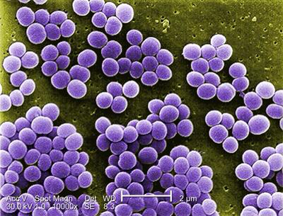
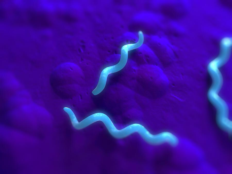
Bacteria are single-celled, prokaryotic microorganisms. Some bacteria are beneficial to humans while others are pathogenic, but a majority of bacteria are harmless to humans. Pathogenic bacteria are responsible for a variety of diseases including strep throat and tetanus. Bacteria come in 3 shapes: coccus(circular), bacillus (rod-shaped), and spirillum (spirally). Bacteria originate from the single-celled organisms that were the first to inhabit the Earth.
- Note: The bacterial shapes are only necessary to remember for division C, although it is always helpful to know them.
Organisms are often classified by their source of energy and source of carbon. Bacteria may be photoautotrophic, utilizing photosynthesis to produce food and oxygen and using carbon dioxide as their source of carbon. This category includes purple and green sulfur bacteria. They may also be chemoautotrophic, making food using the energy from chemical reactions and using carbon dioxide as their source of carbon - these bacteria serve an important role in the nitrogen and sulfur cycles. The two other types are photoheterotrophs (including purple and green non-sulfur bacteria) and chemoheterotrophs (including most bacteria, animals, fungi, and protozoa). Motile bacteria may utilize rotating flagella to move, or they may secrete slime to slide around like a slug. Bacteria may also be nonmotile.
Archaea
Archaea are a group of single-celled microorganisms that were previously thought to be bacteria. Archaea are prokaryotes. Their origin and potential for causing disease are currently unclear; however, archaea are thought to be ancestors of eukaryotes or very close descendants because of their many similarities, including genes and inclusion of enzymes in translation and transcription processes. Unlike bacteria, no known species of archaea form spores.
Archaea are capable of living in extreme habitats and anaerobic environments. They are extremely tolerant to heat, acid, and toxic gases. Archaea are variously involved in the carbon and nitrogen cycles, assist in digestion, and can be used in sewage treatment. They are not known to cause any human diseases.
Fungi
Fungi are eukaryotic organisms that can be single-celled or multi-celled. Fungi have cell walls composed of chitin, unlike the cellulose walls of plants. Fungi are heterotrophic and do not have chloroplasts like photoautotrophs. They grow best in slightly acidic environments and can grow in areas of low moisture. Technically, fungi are more closely related to animals than they are to plants and likely shared a common ancestor with animals. Fungi are responsible for diseases such as athlete's foot. Baker’s yeast (a fungi) is used for bread and brewing. Some fungi are used for antibiotics and others are important decomposers in the ecosystem.
Protists
Protists are eukaryotic but do not have specialized tissues. Algal protists are similar to plants and can go through photosynthesis, but do not have cuticles that prevent water loss. As a result, algal protists must live in water. Animal-like protists are called protozoa and are eukaryotic and heterotrophic. These protists consume other protists and bacteria for food. Some have two nuclei: the macronucleus and the micronucleus. Many move with cilia, flagella, or pseudopodia (in the case of amoebae). They also have complex life cycles. For example, they may exist in a trophozoite, or feeding, form. They can also change into a dormant form known as a cyst, which can help in reproduction.
Endosymbiotic Theory
Championed by Lynn Margulis in the 1960s, the endosymbiotic theory holds that mitochondria and chloroplasts in eukaryotic cells originated from proteobacteria and cyanobacteria, respectively. Evidence for this theory includes that mitochondria and chloroplasts divide through binary fission, not mitosis like the rest of the cell. These organelles, which are the same size as bacteria, also have their own different, circular DNA, their own ribosomes, and two membranes. The two membranes have different chemical compositions, with the outer being similar to the eukaryotic plasma membrane and the inner being similar to bacterial membranes. Chloroplasts in some algae have cell walls of peptidoglycan.
Microbial Growth Curve

When microbes (bacteria) are put in a favorable environment, they usually follow a characteristic pattern of growth. This is represented using the microbial growth curve.
Lag Phase
- During the lag phase of the microbial growth cycle, cells are maturing for doubling; synthesis of RNA, enzymes, etc. Therefore, lag phase in the microbial growth curve is represented by the initial horizontal line.
Exponential Growth Phase
- During the exponential growth, the microbial population undergoes constant doubling. The more "favorable" conditions are, the longer the slope will be, and the faster the growth, the steeper the slope. This is the phase which generation time can be calculated as well.
Stationary Phase
- The stationary phase is another flat portion of the microbial growth curve where it appears that there is no significant increase in the number of cells; showing a stabilization of the population. This is due to the fact that the amount of dying cells is equal to the amount of new cells.
Decline or Death Phase
- Unless the microbes have an infinite source of nutrients, the microbes begin to use up all of the surrounding resources and the waste products begin to pile up. This leads to an unfavorable environment which causes the amount of dying cells to outnumber the amount of new ones.
Gram Staining
- This topic is only tested in Division C.
Gram staining is a type of differential staining, meaning it separates bacteria into two different groups (Gram-positive and Gram-negative) based on their reactions to the procedure. Because of widely varying responses, Gram staining cannot be performed on archaea.
The first step in the procedure is to heat fix the bacteria; then, those bacteria are stained with crystal violet, the primary stain, for one minute. In an aqueous solution, crystal violet disassociates into CV and Cl ions, which penetrate through the cell wall. CV ions react with negatively charged particles in bacterial cells and stain them purple.
The third step is to apply iodine as a mordant, or trapping agent, for one minute. It reacts with the crystal violet and prevents removal of the purple stain. After the remaining iodine is rinsed away, alcohol decolorizer (sometimes acetone) is added until the primary stain is removed in Gram-negative bacteria because alcohol dissolves the outer membrane. In contrast, Gram-positive bacteria retain the primary stain because it becomes trapped in their thick, multi-layered walls of peptidoglycan.
The final step is to apply safranin (sometimes basic fuchsin) as a counterstain. This gives the Gram-negative bacteria their final red-pink color.
Characteristics of Gram-Positive Bacteria
Typically, Gram-positive bacteria produce exotoxins and are susceptible to phenol disinfectants. They retain the blue-purple color of crystal violet in Gram staining because of their thicker walls of peptidoglycan. Unlike Gram-negative bacteria, they lack the periplasmic space between the cytoplasmic and outer membranes because Gram-positive bacteria lack an outer membrane. Certain types of Gram-positive bacilli, most importantly Lactobacilli (used in milk and dairy products), cannot form spores.
Characteristics of Gram-Negative Bacteria
Gram-negative bacteria have thinner walls of peptidoglycan and two membranes and periplasmic space between them. Because of the safranin counterstain, they become red-pink after Gram staining. There are many Gram-negative aerobic (oxygen-using) bacteria.
Types of Microscopes
This requires cleanup to meet the wiki's quality standards. |
Main article: Microscope
- Optical Microscopes: Optical microscope uses visible light (or UV light in the case of fluorescence microscopy) to sharply magnify the samples. The light rays refract with optical lenses. Dating back to the first microscopes that were invented, it is found that they belonged to this category. Optical microscopes can be further subdivided into several categories:
- Compound Microscope: The compound microscope is built of two systems of lenses for greater magnification (an objective and an ocular: eyepiece). The utmost useful magnification of a compound microscope is about 1000x.
- Stereo Microscope (dissecting microscope): The stereo microscope is an optical microscope which magnifies up to about maximum 100x and provides a 3-dimensional view of the specimen. Stereo microscopes are highly useful for observing opaque objects.
- Confocal Laser scanning microscope: Unlike compound and stereo microscopes, Confocal Laser scanning microscopes are reserved for research organizations. Such microscopes are able to scan a sample also in depth. A computer can then assemble the data to create a 3D image.
- Electron Microscopes: Electron microscopes are the most advanced microscopes used in modern science. The electron microscopes essentially function on the principle of a beam of electrons that strikes any objects that comes to its path to magnify it. Electron microscopes are designed specifically for studying cells and small particles of matter, as wells as large objects.
- Scanning Electron Microscope: Scanning Electron Microscope is characterized as a microscope that has lower magnifying power but can provide 3-dimensional viewing of objects. The Scanning Electron Microscope captures the image of the object in black and white after being stained with gold and palladium.
- Reflection Electron Microscope: Reflection electron microscopes are also designed on the principle of electron beams but they are characteristically different from transmission and scanning electron microscopes being that it is built to detect electrons that have been scattered elastically.
- X-ray Microscope: An X-ray microscope uses a beam of x-rays to create an unparalleled high-resolution 3D image. Due to the small wavelength, the image resolution is higher as compared to optical microscopes. The greatest useful magnification is therefore also higher and it lies between the optical microscopes and electron microscopes. X-ray microscopes hold significant importance in science and research and have got one special advantage over electron microscopes- it allows observing the structure of the living cells. It is adept at slicing together thousands of images to generate a single 3D X-ray image.
- Scanning Helium Ion Microscope (SHIM or HeIM): Scanning Helium Ion Microscope is a new imaging technology which uses a beam of Helium ions beams to generate an image. This technology has several advantages over the traditional electron microscopes; one advantage lies in the fact that the sample is left mostly intact (due to the low energy requirements) and that it provides a high resolution. The first commercial systems were released in 2007.
- Scanning acoustic microscope (SAM): Scanning acoustic microscope uses focused sound waves to generate an image. An acoustic microscope has a wide range of applications in materials science to detect small cracks or tensions in materials. The scanning acoustic microscope is a powerful tool which can also be used in biology to study the physical properties of the biological structure and help uncover tensions, stress and elasticity inside the biological structure.
- Electron Microscopes: Modern electron microscope uses accelerated electrons and can magnify up to 2 million times due to the very small wavelength of high energy electrons. The high energy electrons as a source of illumination are quite tough on the sample being observed. Because of the much shorter wavelength, the electron microscope has a higher resolving power than a light microscope. To reveal the structure of objects, it may initially acquire a long time to completely dehydrate and prepare the specimen. A sleek layer of a metal could be on the other hand used to coat some of the biological specimens for easy observation.
- Neutron Microscope: Still under an experimental stage, Neutron microscope generates a high-resolution image and may offer better contrast than other forms of microscopy. The new technology would use neutrons instead of beams of light or electrons to generate high-resolution images.
- Scanning Probe Microscopes: Scanning Probe Microscope helps visualize individual atoms. The image of the atom is computer-generated, however. It provides the researchers with an imaging tool for the future where a small tip measures the surface structure of the sample. These specialized microscopes provide high image magnification to observe three-dimensional specimens. If an atom projects out of the surface, then a higher electrical current flows through the tip. The amount of current that flows is proportional to the height of the structure. A computer then assembles the position data of the tip. An enhanced 3D image is generated.
National Topics
This section is incomplete. |
This section requires cleanup to meet the wiki's quality standards. |
On occasion, the rules will specify certain topics to only be tested at the national level. These are discussed below.
Microbial Population Explosions
The most common example of a microbial population explosion is an algal bloom/red tide, an an aggregation of red dinoflagellates in an aquatic ecosystem. Fertilizer-rich runoff, often from a farm, is the typical cause for an algal bloom.The abundance of nutrients in the fertilizer becomes a part of the runoff and enters a nearby body of water along with it. This eutrophication fuels algal growth, allowing dinoflagellates to reproduce rapidly.
As a result, certain algae can produce potentially lethal toxins that are harmful to animals. Algal blooms are also expensive to treat, greatly impacting water treatment plants. Perhaps the most devastating consequence of all is ocean dead zones. Given a massive population of algae, many will eventually die due to lack of space, buildup of toxins, etc. Marine decomposers (i.e. bacteria) break down the organic material of the dead dinoflagellates, which requires oxygen. Oxygen in surrounding waters is depleted, causing hypoxia and eventually anoxia (the state of having no oxygen). This causes most of the marine life to die, hence the name "ocean dead zone".
Microbial population explosions can also be influenced by their behavioral preferences. For example, fungi grow best at a slightly acidic pH. One could go even further and look at the diversity of behaviors in bacteria alone. Neutrophiles are most comfortable at a neutral pH of 7, which tends to be suitable for most pathogens. Obligate anaerobes cannot tolerate any oxygen and die in its presence, making their environment anoxic. There are also xerophiles, which thrive in extremely dry surroundings. The adaptations of microbes are very broad, which allows them to rapidly multiply in almost any given environment.
Microbial Competition and Communication
Microbial communication is done through quorum sensing (qs). After attaching to a surface, qs bacteria send out autoinducers that indicate the population of nearby bacteria. If there are enough bacteria nearby, it starts creating a biofilm. Just like all organisms, microbes compete to survive. Some microbes "stab and poison" neighboring cells to kill them. Others colonize an excess amount of space to prevent other bacteria from growing there. Many methods of competition exist, though they can all be divided into two types: scramble competition and contest competition.
In scramble competition, participants use up as many available resources as possible to prevent other organisms from using them. To visualize this, imagine a ton of children are standing on a field and someone throws out a ton of quarters. The children would try to get the most quarters that they can to stop others from getting them. Contest competition is just like it sounds: direct competition between species, with the winner getting exclusive access to the resources. An analogy for this is the same scenario, except the quarters aren't thrown out and are instead reserved. All the kids are given weapons, and the last person standing gets the quarters, similar to the Hunger Games.
In every competition however, there are cheaters. The same goes for the microbial world, where "social cheaters" exist. An example of this is bacteria that mimic P. aeruginosa, which happens to produce a certain autoinducer that attracts bacteria. Once gathered, they produce a large quantity of antimicrobials to eliminate competitors. Other bacterias can analyze this behavior and replicate it without undergoing evolution.
Microbiomes
Biofilms
Diseases for 2017-2018
For specific information about each disease, see Microbe Mission Diseases List
Viral Diseases
Viral diseases are immune to antibiotics; therefore, vaccines are typically the best form of prevention.
- AIDS (caused by the HIV virus)
- Chicken Pox and Shingles (caused by the varicella zoster virus)
- Common cold (often caused by the rhinovirus but can also be caused by coronaviruses and adenoviruses)
- Dengue fever (caused by the mosquito-borne dengue virus)
- Ebola Haemorrhagic Fever (caused by the ebola virus)
- Hepatitis
- Influenza (caused by viruses of the family Orthomyxoviridae)
- Measles (caused by the morbillivirus)
- Mumps (caused by the mumps virus)
- Mononucleosis, also known as mono or glandular fever (caused by the Epstein-Barr virus)
- Norovirus
- Polio (caused by the poliovirus)
- Rabies (caused by the rabies virus)
- Rubella
- Yellow Fever
- Zika
Bacterial Diseases
Bacterial diseases may often be treated by antibiotics.
- Anthrax
- Botulism
- Cholera
- Chlamydiasis
- Dental Caries (tooth decay)
- Legionnaire's Disease
- Lyme Disease
- MRSA
- Peptic Ulcer Disease
- Pertussis (whooping cough)
- Rocky Mountain Spotted Fever
- Strep throat
- Syphilis
- Tetanus
- Tuberculosis
Fungal Diseases
The technical term for a fungal disease is a mycosis.
- Athlete's foot
- Dutch Elm Disease
- Histoplasmosis
- Potato Blight (caused by Alternaria solani)
- Ringworm
- Thrush
- White Nose Syndrome
Protozoan & Algal Diseases
- Malaria
- Paralytic Shellfish Poisoning
- Naegleria
- Giardiasis
- Cryptosporidiosis
Prionic Diseases
- Chronic Wasting Disease
- Kuru
Parasitic Worms
- Hookworm
- Pinworm
- Schistosomiasis
- Tapeworm
- Trichinosis
National Tournament Added Diseases/Important Genera
- Pseudomonas aeruginosa
- Wolbachia
Sample Exercises
1. Provide two differences between bacteria, viruses, and fungi.
2. Using the following key, determine (from pictures) which cell, A, B, or C is considered an alga.
3. Based on the following graph, determine which organism is best suited for growth in acid environments.
4. A cell is observed through a light microscope at 4x magnification. The cell takes up about half of the visual field. What is the approximate length of this organism?
5. Students observe a Petri plate with many different colonies on it. Based on the color of the colony, how many different kinds of organisms do you detect? Which type of organism appears to be the most prevalent?
6. From the following picture, identify the organelle, provide its function, and state which type of microbe it is unique to.
7. What type of microbe is involved in the production of most breads? What type of organism is responsible for polio?
8. Based on the following graph, what will be the microbial population/ml after 3.5 hours of growth?
9. Provide two distinctive properties of viruses, then provide the name of two diseases that are caused by viruses. As a variation on this type of question, match the disease with the type of microbe that causes it.
10. Match each microbe that causes the following disease: Rubella, Schistosomiasis, Ebola, Scrapie, Malaria.
Useful links
- Microbe Mission page on soinc.org
- Has information specific to cells -- Has a lot of useful information -- Use the links on that page too!
- Has vocabulary
- Bacteriology text
- Microbiology text
- CellCraft -- a game which helps you to learn the basic organelles of a cell.
- Microbiology Online -- a great place to find in-depth information on cell organelles